
Last week, I looked into some breakthroughs in the history of veterinary medicine and technology and how these technologies work. I did research on stem cell therapy, anesthesia and anesthetic monitoring, and 3D printing. This week, I want to continue on the same topic, looking into more breakthroughs such as laser surgery and the different types of diagnostic imaging.
Laser surgery/therapy
-Laser surgery is no longer just for human corrective eye surgery (1). It is a type of treatment that has been around for decades, but only recently has it become common in veterinary medicine (2).
-“Laser” is actually an acronym that stands for “light amplification by stimulated emission of radiation,” and it is a device that emits radiation through a flow of photons of light energy. Laser light is monochromatic, coherent and collimated. This means that it has one wavelength, all the photons move in the same phase and direction, and the laser beam has very little divergence over a distance (light does not spread out). These properties let therapy laser lights to focus on very specific areas, penetrate skin without heating or damaging it, and interact with tissue with minimal side effects. Wavelength also changes how deep it penetrates, with longer wavelengths penetrating deeper into the tissues. Shorter wavelengths can be used to treat superficial wounds and joint injuries, while muscle injuries should generally be treated with longer wavelengths (2).
These concentrated sources of light may be used for many procedures including:
- spaying and neutering (1)
- declawing (1)
- skin wounds (2)(6)
- tendon and ligament injuries (2)
- muscle injuries (2)
- injuries to the nervous system or neurologic conditions (2)
- osteoarthritis (2)(3)
-Surgical lasers can be extremely beneficial because it causes no hemorrhaging (or very little), which gives the veterinarian a clear operative field, it reduces pain and swelling, and it is more precise. Over the past several years, there have been much more higher-powered lasers available, and the higher laser power allows veterinarians to cut at the same speed as they would with a scalpel blade, but with the added benefits that surgical lasers provide (4).
–Moreover, the design of these modern surgical lasers has improved drastically in recent years. The flexible fiber CO2 laser delivery system has tip-less handpieces that are light and like a pen, which is far less stressful to use than the older arm systems that are bulky and use technology from the 1980’s (4).
-The most commonly used lasers in veterinary practice are the carbon dioxide and diode lasers. Usually, the carbon dioxide lasers are more practical for veterinary practice, except for endoscopy procedures (4) (which are non-surgical). In that case, the fiber-based diode laser would be more practical (4).
-However, a drawback to laser surgery is the cost. They may have dropped in price in recent years, but they are still expensive and may not be worth it for all veterinary practices. Using these units may add an additional 50 to 100 dollars or more to the cost of a surgery (5).
Diagnostic Imaging
-Veterinary diagnostic imaging includes many imaging methods such as radiographs (x-rays), ultrasound, MRIs and CT scans, which are mostly non-invasive and entirely painless. However, some methods may need sedation or anesthesia due to the animal needing to be kept still for adequate images to be made. These images are used to collect information on the animal to help veterinarians in making a medical or even surgical plan (8).
Radiography (X-rays)
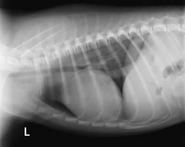
(13)
-X-rays have been used for decades to create images known as radiographs (black, white and grey images), and it is the most commonly used imaging procedure in veterinary practices. X-rays are good for producing images of bones, foreign objects, and large body cavities. They are frequently used for detecting fractures, tumors, injuries, infections and deformities. Although they might not always provide enough information to determine the exact cause of the problem the animal has, they can help the veterinarian in determining which other tests that might be needed to make a proper diagnosis (9)(10).
-However, the tissues are soft and make it difficult to see them. Therefore, veterinarians use specialized x-ray techniques, known as contrast procedures, to help give more detailed images of body organs. Basically, they give the animal a dye that will block x-rays. This can be done intravenously to look at organs such as the kidneys or heart, or it can be done by mouth to look at the digestive tract. The x-rays taken after the dye is given to the animal will then outline the organs where the dye collected, which makes it easier to find any abnormalities (9)(10).
For example, when I went to the veterinary clinic my mom works at for “Take Your Kid to Work Day” in grade 9, there was an ill dog, and it was suspected that it had swallowed something it wasn’t supposed to have swallowed. Therefore, they fed the dog barium, a white substance, and performed several x-rays. Looking at the images after this, we could see a white blockage (highlighted by the barium) in its digestive tract that had not moved along after performing more x-rays hours later, so the veterinarian knew she had to do a surgery to remove whatever was in its digestive tract.
Initially, radiographs were produced on film; however, recent technology advances have allowed them to be produced and stored digitally (9)(10)(11)(12)(13). It was in 2004 that the Diagnostic Imaging service upgraded from film to computed radiography (CR), which used a cassette system, and it was in 2010 that they added digital radiography, which could process images in seconds and produce high quality diagnostic images with less radiation exposure and a higher efficiency (13).
Fluoroscopy
-Fluoroscopy is a type of radiographic imaging that does a continuous x-ray so that it shows real-time moving body structures on a monitor (11)(12). It is usually used for surgical and cardiac procedures because it lets the veterinarian visualize the internal location of catheters and other instruments when they are inserted into the animal’s body (12).
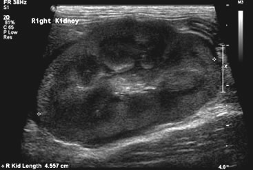
Ultrasonography
-Ultrasonography (ultrasound) uses ultrasonic sound waves to produce images of body structures using the patterns of echoes reflected off the tissues and organs (9)(10)(11)(12)(14). Ultrasound scans are able to show the size and shape of organs and any abnormalities they might have. They are both painless and non-invasive, and don’t pose any risk of complications (9)(10).
(14)
Nuclear Scintigraphy
-With nuclear scintigraphy, the animal is dosed with an element that emits a type of radiation called gamma rays. The element is detected in the body using a special gamma camera attached to a computer, which produces the image. The element is connected to a molecule that has an affinity for the organ or tissue they are examining. If the organ or tissue metabolize the molecule or if the molecule remains for only a short period of time (9)(10), consecutive images may be used to evaluate the function of the organ or tissue (9)(10)(15).
Computed Tomography (CT)
-Computed Tomography (CT) is a type of x-ray procedure that is computer-enhanced, and it is used for detecting abnormalities in body organs (9)(10)(11). The animal lies on a motorized bed inside of a CT scanner, which takes x-rays from many different angles, and when one scan is finished, the bed moves forward and another one is taken. A computer then uses these scans to create cross-sectional images of the body part being examined and displays them on a monitor, and giving the animal a dye intravenously will make it easier to spot any abnormalities (9)(10). CT scans can be used to detect structural changes deep inside the body, such as tumors (9)(10)(11). Because the animal needs to stay still for quite a while during this process, they are anesthetized (9)(10).
Magnetic Resonance Imaging (MRI)
-Magnetic Resonance Imaging (MRI) is now an alternative to CT scans. With MRIs, an extremely strong magnetic field produces detailed anatomic images. X-rays are not used in this procedure, and it is very safe. However, due to the cost and size of the equipment and the need for specially trained technicians, it isn’t used very often for pets (9)(10). For this procedure, the animal is put into a tubular electromagnetic chamber and pulses of radio waves are directed at it, causing the body tissue to emit radio frequency waves, which are then converted into an image with a computer. Because the procedure takes a long time and the animal cannot move, anesthesia is often used (9)(10)(16).
That’s all for now! In my next round of research, I plan to do research on how veterinary medicine and technology has benefited not just animals, but humans and society in general too. If you have any ideas, suggestions or websites I could use, feel free to leave a comment. Thanks for reading!
Sources:
(1) https://www.vettech.org/25-amazing-veterinarian-breakthroughs-in-the-last-10-years
(2) https://ivcjournal.com/laser-therapy-veterinary-medicine/
(3) https://ivcjournal.com/laser-therapy-for-osteoarthritis/
(4) https://www.veterinarypracticenews.com/a-look-at-veterinary-lasers/
(5) https://animalwellnessmagazine.com/new-veterinary-advancements/
(6) https://vetprise.com/blog/advances-veterinary-medicine/
(8) https://www.harlingenveterinaryclinic.com/services/dogs/canine-ultrasound-mri-x-rays-medical-imaging
(9) https://www.msdvetmanual.com/special-pet-topics/diagnostic-tests-and-imaging/diagnostic-imaging
(10) https://www.merckvetmanual.com/special-pet-topics/diagnostic-tests-and-imaging/diagnostic-imaging
(11) https://vetmed.oregonstate.edu/diagnostic-imaging-techniques
(12) https://www.dispomed.com/career-spotlight-veterinary-diagnostic-imaging-specialist/
(16) https://canadawestvets.com/library/magnetic-resonance-imaging-mri/
Hi Jessica,
This was a very informative post! I was quite surprised to read that laser surgery is now being used in veterinary medicine and has been for awhile. With this new technology and the growing use of laser surgery, do you think that laser surgery might take away the need for a doctor to use a scalpel altogether? Also, besides cost, could there be any other drawbacks or risks to using laser surgery?
I saw that for your next post, you plan to look into the benefits veterinary medicine has beyond animals, by helping humans and our community. Here are some websites that could help with your next round:
This website looks at the role veterinarians play beyond the work they do to help animals. It looks at how Veterinarians help protect the environment, research new things etc.:
https://www.avma.org/public/YourVet/Pages/Veterinarians.aspx
http://eara.eu/en/animal-research/veterinary-medicine/
Good luck on your next round!!
– Madison Ciulla